Heat
DHT27 - Thermal Imager - Fluke Ti10
Objective:
- To demonstrate the principle of thermal imaging
Apparatus:
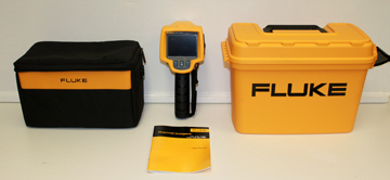
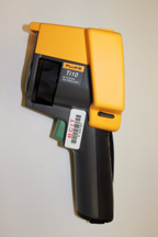
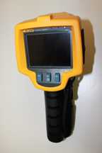
- Thermal Imager - Fluke Ti10 and accessories (-20 degrees Celsius to + 250 degrees Celsius)
Thermal Imaging
Here's how thermal imaging works:
- A special lens focuses the infrared light emitted by all of the objects in view.
- The focused light is scanned by a phased array of infrared-detector elements. The detector elements create a very detailed
temperature pattern called a thermogram. It only takes about one-thirtieth of a second for the detector array to obtain the
temperature information to make the thermogram. This information is obtained from several thousand points in the field of view
of the detector array.
- The thermogram created by the detector elements is translated into electric impulses.
- The impulses are sent to a signal-processing unit, a circuit board with a dedicated chip that translates the information from the elements into data for the display.
- The signal-processing unit sends the information to the display, where it appears as various colors depending on the intensity of the infrared emission. The combination of all the impulses from all of the elements creates the image.
Method:
- see the Fluke Ti10 manual for operating instructions